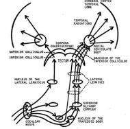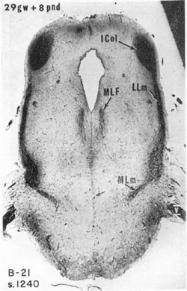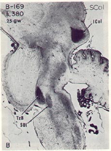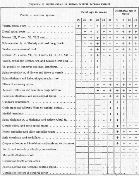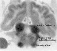
| Figure11 is a diagram of the auditory pathway from the cochlear nerve in the lower brainstem to the temporal lobes of the cerebral cortex. The inferior colliculi are the posterior pair of "hills" (Latin colliculi) in the midbrain tectum. Figure 12 is an autoradiogram from the investigations of Landau et al. (1955) on cerebral blood flow [1]; it shows the greatest uptake of radioactive tracer in the inferior colliculi. Figure 12 can be compared with figure 11 for location of the inferior colliculi, the superior olives, and the lateral lemniscal tracts connecting these important way stations in the brainstem auditory pathway. The dramatic autoradiographic picture in figure 5 is from a paper by Kety (1962), who was director of the group that first used radioactive tracers to study blood flow in the brain [2]. Figure 6 from observations of myelination by Yakovlev and Lecours (1967) shows that the greatest degree of myelin development in a human fetus at 29 weeks gestation is in the same structures with highest blood flow, the inferior colliculi and the lateral lemniscal tract connections to it from the superior olives [3]. Myelination is an important measure of maturation, and the study by Yakovlev and Lecours confirmed earlier research by Langworthy (1933) who also found the auditory system of the brain among the earliest to mature [4]. Langworthy's data is shown in figure 7. Confirmation of early myelination and function of the auditory system can be found in the paper by Moore et al. (1995) [5]. Early maturation of the auditory system suggests its possible involvement in stimulating growth of later developing areas of the cerebral cortex. Growth of the auditory receptive areas of the temporal lobes and speech motor areas in the frontal lobes are likely dependent at least in part on neurotransmissions from the brainstem auditory system. Neurotransmission does not necessarily require acoustic stimulation; development of the temporal and frontal lobes takes place normally in children born deaf, but their deafness is due to impairment within the lower auditory system, not the inferior colliculi and thalamic auditory nuclei. |