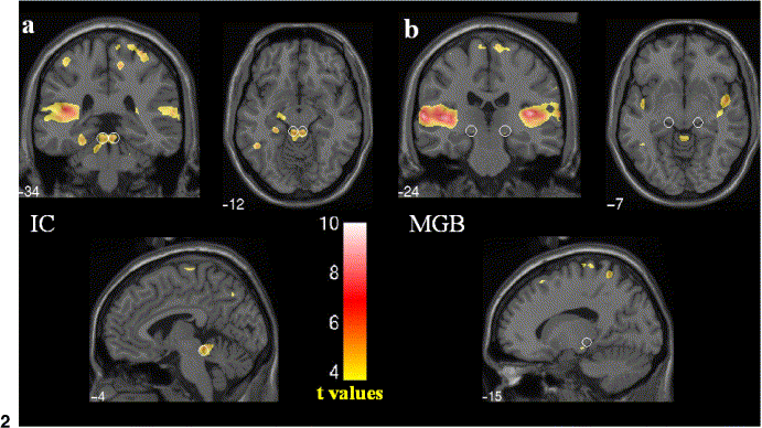
Figure 5 – Visualization of inferior colliculi by fMRI
(a) Three orthogonal slices (coronal, horizontal, sagittal) centered on the anatomically
defined inferior colliculi (IC) - white circles superimposed on an individual T1 image.
(b) Images as in (a) but with slices centered on the anatomical center of the medial
geniculate bodies MGB (from Budd et al. 2003).
back