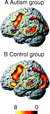
Figure 8 – Language areas in autism
Figure 8: Brain activation of autistic (A) and control (B)
groups (Sentence versus Fixation contrast). Autistic
participants show less activation in the left inferior
frontal gyrus (LIFG) than the control group, but more
activation in the left posterior superior temporal gyrus
(LSTG) than the control group.
back