2 - Metabolic activity in the brain
The cerebral cortex is not the most metabolically
active part of the brain; this is not intuitive and may
not seem to make sense. The cerebral cortex can
be seen on functional MRI images (fMRI) to be
selectively activated in areas responsive to
particular sensory stimuli. Activity at a continuously
high level is not needed in the cortex, anymore than
computer memory locations need to be constantly
active – they are activated only when accessed to
store or retrieve data.
The brainstem nuclei damaged by asphyxia at birth
are metabolically the most active centers of the brain
[1, 2]; they may function as multiplexing gates that
handle the flood of sensory stimuli to which we are
constantly exposed. As Fisch (1970) noted, the
auditory system is continuously active, even while we
sleep, and remains constantly vigilant of what goes
on in our environment [3]. Sokoloff (1981)
concluded from measurements of glucose uptake
that “the inferior colliculus is clearly the most
metabolically active structure in the brain” [2].
During a period of asphyxia such as that inflicted in
the experiments with newborn monkeys, brainstem
nuclei sustain damage; the cerebral cortex is not
immediately affected [4]. Myers (1972) found that
partial hypoxia or circulatory insufficiency late in
gestation is damaging to the cortex, and produces
the pattern of damage responsible for cerebral palsy
[5]. Protective mechanisms go into action during
periods of oxygen insufficiency that preserve the
activity in the brainstem areas of high metabolic rate;
the cortex then becomes vulnerable.
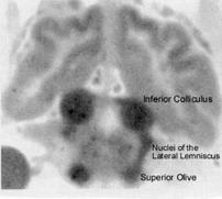
Figure 6 is an autoradiogram from the experiments
on cerebral blood flow in cats. It was published in an
article by Seymour Kety, who was the principal
investigator of these experiments [6]. The blood flow
data from the same research is shown in table 3.
- Landau WM et al. (1955) The
local circulation of the living
brain; values in the
unanesthetized and
anesthetized cat.
- Sokoloff L (1981) Localization
of functional activity in the
central nervous system by
measurement of glucose
utilization with radioactive
deoxyglucose.
- Fisch L (1970) The selective
and differential vulnerability of
the auditory system.
- Windle WF (1969) Brain
damage by asphyxia at birth.
- Myers RE (1972) Two
patterns of perinatal brain
damage and their conditions
of occurrence.
- Kety SS (1962) Regional
neurochemistry and its
application to brain function.
The highest blood flow
and metabolism in the brain is
in the brainstem nuclei of the
auditory pathway.
Landau et al (1955) used a
radioactive tracer to investigate
cerebral blood flow in laboratory
animals [1]. The picture to the
left is an autoradiogram of the
brain of a cat 60 seconds after
injection of a tracer. It shows
the greatest perfusion (thus
greatest blood flow) in the
inferior colliculi, superior olives,
and lateral lemniscal tracts
connecting these relay nuclei in
the brainstem auditory pathway.
Autoradiogram picture from Kety (1962) and the
research of Landau et al. (1955), with permission
from Columbia University Press.
Figure 6
- Landau WM, Freygang WH, Rowland LP, Sokoloff L, Kety SS (1955) The local circulation
of the living brain; values in the unanesthetized and anesthetized cat. Transactions of
the American Neurological Association 1955-1956;(80th Meeting):125-129.
- Sokoloff L (1981) Localization of functional activity in the central nervous system by
measurement of glucose utilization with radioactive deoxyglucose. Journal of Cerebral
Blood Flow and Metabolism 1:7-36.
- Fisch L (1970) The selective and differential vulnerability of the auditory system. In GEW
Wolstenholm and J Knight, (Eds), Sensorineural Hearing Loss: A Ciba Foundation
Symposium (pp 101-116). London: Churchill.
- Windle WF (1969) Brain damage by asphyxia at birth. Scientific American 221(#4):76-
84.
- Myers RE (1972) Two patterns of perinatal brain damage and their conditions of
occurrence. American Journal of Obstetrics and Gynecology 112:246-276.
- Kety SS (1962) Regional neurochemistry and its application to brain function. In French,
JD, ed, Frontiers in Brain Research. New York: Columbia University Press, pp 97-120.
Table 3: Cerebral blood flow data in cats, using a
radiographic tracer (from Landau et al. 1955)
|
Brain Structure
Brain System
Flow Rate
(cc/gm/min)
Auditory
Auditory
Visual
Visual
Subcortical motor
1.80
1.38
1.30
1.25
1.22
1.21
1.15
1.10
1.03
0.88
0.87
0.24
0.23
0.14
Inferior colliculus
Sensory-motor cortex
Auditory cortex
Visual cortex
Medial geniculate
Lateral geniculste
Superior colliculus
Caudate nucleus
Thalamus
Association cortex
Cerebellar nuclei
Cerebellar white matter
Cerebral white matter
Spinal cord white matter
Highest blood flow in the brain
is to the inferior collicui