The same nuclei in the
brainstem auditory pathway that
are first to become myelinated
also have the highest blood flow
and metabolism.
|
3 - Site of highest metabolic activity
Sokoloff et al. (1977) refined the autoradiographic
method further by using carbon-14 labeled
deoxyglucose [1]. This is an analogue of glucose that
enters the brain but then is not further metabolized.
Results of this method provide a measure of glucose
utilization that can differ from blood flow in some
circumstances. Normally glucose uptake is greatest in
the same brain areas with the highest rate of
circulation. Baseline values for blood flow and
deoxyglucose uptake can both be used as estimates
of metabolic rate, and these methods are now
revealing more and more the often surprising effects
of drugs and other factors that alter homeostasis in
the brain.
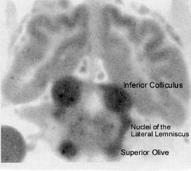
Sokoloff (1981) discussing measurements of regional
glucose uptake stated, “the inferior colliculus is clearly
the most metabolically active structure in the brain”
[2]. Figure 12 is an autoradiographic photo (from an
article by Kety 1962) that shows the high rate of blood
flow in the inferior colliculus [3].
Using other measures of cerebral metabolism,
Rahner-Welsch et al (1995) and Zeller et al (1997)
reported the highest levels of glucose transport
proteins in the inferior colliculus of rat brains [4, 5].
Hovda et al (1992) found the mitochondrial enzyme
cytochrome oxidase (COX) to be highest in the inferior
colliculus of developing cats, corresponding to the
finding of Chugani et al (1991) of greatest uptake of
glucose during early development [6, 7]. Gonzalez-
Lima (1997) confirmed the high levels of COX in the
inferior colliculus in the human brain, and that this
enzyme is diminished in the brains of Alzheimer
patients [8].
Energy production in the brain supports
neurotransmission, and neurons that are involved in
the most active circuits of the brain also have the
greatest need for metabolic maintenance.
Transcription of DNA to messenger and transfer RNA
components, then assembly of peptide subunits for
enzymes that synthesize neurotransmitters and
accomplish aerobic energy production are on-going
processes that must take place at high levels. Kirikae
et al. (1988) demonstrated a coupling of glucose
metabolism to the production of protein and
maintenance of structural components of nerve cells.
The rate of incorporation of the amino acid valine into
proteins was found to be highest in the inferior
colliculus, as expected from the high rate of energy
production in this auditory nucleus [9].
- Sokoloff L, Reivich M, Kennedy C, Des Rosiers MH, Patlak CS, Pettigrew KD, Sakurada O,
Shinohara M (1977) The [14C]deoxyglucose method for the measurement of local cerebral
glucose utilization: theory, procedure, and normal values in the conscious and anesthetized
albino rat. Journal of Neurochemistry 28:897-916.
- Sokoloff L (1981) Localization of functional activity in the central nervous system by
measurement of glucose utilization with radioactive deoxyglucose. Journal of Cerebral
Blood Flow and Metabolism 1:7-36.
- Kety SS (1962) Regional neurochemistry and its application to brain function. In French, JD,
ed, Frontiers in Brain Research. New York: Columbia University Press, pp 97-120.
- Rahner-Welsch S, Vogel J, Kuschinsky, W (1995) Regional congruence and divergence of
glucose transporters (GLUT1) and capillaries in rat brains. Journal of Cerebral Blood Flow
and Metabolism 15:681-686.
- Zeller K, Rahner-Welsch S, Kuschinsky W (1997) Distribution of Glut1 glucose transporters
in different brain structures compared to glucose utilization and capillary density of adult rat
brains. Journal of Cerebral Blood Flow and Metabolism 17:204-209.
- Hovda DA, Chugani HT, Villablanca JR, Badie B, Sutton RL (1992) Maturation of cerebral
oxidative metabolism in the cat: a cytochrome oxidase histochemistry study. Journal of
Cerebral Blood Flow and Metabolism 12:1039-1048.
- Chugani HT, Hovda DA, Villablanca JR, Phelps ME, Xu, W-F (1991) Metabolic maturation of
the brain: a study of local cerebral glucose utilization in the developing cat. Journal of
Cerebral Blood Flow and Metabolism 11:35-47.
- Gonzalez-Lima F, Valla J, Matos-Collazo S (1997) Quantitative cytochemistry of cytochrome
oxidase and cellular morphometry of the human inferior colliculus in control and Alzheimer's
patients. Brain Research 752:117-126.
- Kirikae M, Diksic M, Yamamoto YL (1988) The transfer coefficients for L-valine and the rate
of incorporation of L-[1-14C] valine into proteins in normal adult rat brain. Journal of
Cerebral Blood Flow and Metabolism 8:598-605.
| Deoxyglucose uptake in monkey & rat brain |
| Brain Structure | Monkey | Albino Rat | Brain System |
| SD 1-4 | SD 2-7 |
| Inferior colliculus | 103 | 197 | auditory |
| Auditory cortex | 79 | 162 |
| Vestibular nucleus | 66 | 128 |
| Medial geniculate | 65 | 131 | auditory |
| Superior olivary nucleus | 63 | 133 | auditory |
| Visual cortex | 59 | 107 |
| Mammillary body | 57 | 121 | limbic |
| Superior colliculus | 55 | 95 | auditory |
| Thalamus, lateral nucleus | 54 | 116 |
| Caudate-putamen | 52 | 110 | subcortical motor |
| Cochlear nucleus | 51 | 113 | auditory |
| Cerebellar nuclei | 45 | 100 |
| Sensorimotor cortex | 44 | 120 |
| Lateral geniculate | 39 | 96 | visual |
| Hippocampus | 39 | 79 | limbic |
| Cerebellar cortex | 31 | 57 |
| Cerebellar white matter | 12 | 37 |
Figure 12 -
Blood flow and metabolism
are not uniform throughout
the brain. The same nuclei
in the brainstem auditory
pathway that are first to
become myelinated during
prenatal life also have the
highest blood flow and
metabolism.
|
 - Sokoloff L et al. (1977) The
[14C]deoxyglucose method
for the measurement of local
cerebral glucose utilization:
theory, procedure, and
normal values in the
conscious and anesthetized
albino rat.
- Sokoloff L (1981)
Localization of functional
activity in the central nervous
system by measurement of
glucose utilization with
radioactive deoxyglucose.
- Kety SS (1962) Regional
neurochemistry and its
application to brain function.
- Rahner-Welsch S et al.
(1995) Regional congruence
and divergence of glucose
transporters (GLUT1) and
capillaries in rat brains.
- Zeller K et al. (1997)
Distribution of Glut1 glucose
transporters in different
brain structures compared
to glucose utilization and
capillary density of adult rat
brains.
- Hovda DA et al. (1992)
Maturation of cerebral
oxidative metabolism in the
cat: a cytochrome oxidase
histochemistry study.
- Chugani HT et al. (1991)
Metabolic maturation of the
brain: a study of local
cerebral glucose utilization
in the developing cat.
- Gonzalez-Lima Fet al. (1997)
Quantitative cytochemistry of
cytochrome oxidase and
cellular morphometry of the
human inferior colliculus in
control and Alzheimer's
patients.
- Kirikae M et al. (1988) The
transfer coefficients for L-
valine and the rate of
incorporation of L-[1-14C]
valine into proteins in
normal adult rat brain.
From Kety (1962) with permission from
Columbia University Press
|