7 – Blood flow in the brain
Myers (1972) cited publications by Kety and Sokoloff
on blood flow in the brain and pointed out, "The
inferior colliculus, the structure most outstandingly
vulnerable to total asphyxia, is also the structure most
highly perfused with blood" [1, p254]. Nevertheless,
Myers did not question why this structure should have
such a high rate of blood flow. On the contrary and on
the same page he states emphatically, "The brainstem
injury pattern produced in the monkey fetus by total
asphyxia bears no relation to the brain pathology
typifying human perinatal damage."
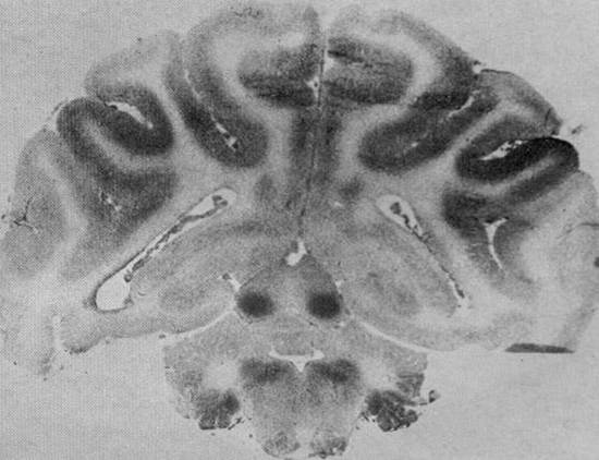
Figure xx is an autoradiograph from Myers' article
showing greatest localization of the radiographic tracer
in the inferior colliculi of a newborn monkey [1, p254].
Myers, unlike Ranck and Windle (1959), did not
mention the neuropathology of kernicterus, but did cite
two symposia presentations (by JB Brierley) reporting
brainstem damage in young children following cardiac
arrest, his own work with Miller on circulatory arrest in
adult monkeys, and a paper describing brainstem
injury caused by exposure to hyperbaric oxygen [4, 5].
According to Myers, it is the infant heart (not the infant
brain) that is resistant to anoxia, "Only the protected
adult heart or the tolerant infant heart is able to
support a sufficient head of blood pressure in the
postarrest period to avert the hemishperal injury
pattern which typifies cerebral malperfusion" (p255).
Preventing damage to the cerebral cortex was of
overriding interest to Myers. For some reason Myers,
in this valuable research study, was unable to interpret
the finding of brainstem damage as being important
enough to investigate further.
Myers (1972) cited publications by Kety and Sokoloff
on blood flow in the brain and pointed out, "The
inferior colliculus, the structure most outstandingly
vulnerable to total asphyxia, is also the structure most
highly perfused with blood" [1, p254]. Nevertheless,
Myers did not question why this structure should have
such a high rate of blood flow. On the contrary and on
the same page he states emphatically, "The brainstem
injury pattern produced in the monkey fetus by total
asphyxia bears no relation to the brain pathology
typifying human perinatal damage."
Figure xx is an autoradiograph from Myers' article
showing greatest localization of the radiographic tracer
in the inferior colliculi of a newborn monkey [1, p254].
Myers, unlike Ranck and Windle (1959), did not
mention the neuropathology of kernicterus, but did cite
two symposia presentations (by JB Brierley) reporting
brainstem damage in young children following cardiac
arrest, his own work with Miller on circulatory arrest in
adult monkeys, and a paper describing brainstem
injury caused by exposure to hyperbaric oxygen [4, 5].
According to Myers, it is the infant heart (not the infant
brain) that is resistant to anoxia, "Only the protected
adult heart or the tolerant infant heart is able to
support a sufficient head of blood pressure in the
postarrest period to avert the hemishperal injury
pattern which typifies cerebral malperfusion" (p255).
Preventing damage to the cerebral cortex was of
overriding interest to Myers. For some reason Myers,
in this valuable research study, was unable to interpret
the finding of brainstem damage as being important
enough to investigate further.
- Myers RE (1972) Two patterns of perinatal brain damage and their conditions of
occurrence. - Ranck JB, Windle WF (1959). Brain damage in the monkey, Macaca mulatta, by
asphyxia neonatorum. - Brierley JB (1965) The influence of brain swelling, age and hypotension upon the
pattern of cerebral damage in hypoxia. In F.Lüthy & A. Bischoff, eds. Proceedings
of the Fifth International Congress of Neuropathology, Zürich, 31 August - 3
September 1965), Excerpta Medica Foundation International Congress Series No
100, Amsterdam.(Harvard/Countway call # ZW 1 E939 no.100 1965). - Brierley JB (1967) Comment on Windle WF, Progressive degenerative changes in
brains of monkeys surviving neonatal asphyxia. In LS James, RE Myers, GE Gaul,
eds. Brain damage in the fetus or newborn from hypoxia or asphyxia, report of the
fifty-seventh Ross Conference on Pediatric Research, Columbus, p 26.
(Harvard/Countway Serial, Report of the Ross Conferences v.30-102,1959-1992). - Miller JR, Myers RE (1972) Neuropathology of systemic circulatory arrest in adult
monkeys. - Balentine JD. (1968) Pathogenesis of central nervous system lesions induced by
exposure to hyperbaric oxygen.
In progress
- Autoradiograph
showing blood flow in the brain
of a newborn monkey, showing
greatest blood flow is to the
inferior colliculi in the midbrain.
showing blood flow in the brain
of a newborn monkey, showing
greatest blood flow is to the
inferior colliculi in the midbrain.
- Myers RE (1972) Two
patterns of perinatal
brain damage and their
conditions of
occurrence. - Ranck JB, Windle WF
(1959). Brain damage in
the monkey, Macaca
mulatta, by asphyxia
neonatorum. - Brierley JB (1965)
- Brierley JB (1967)
- Miller JR, Myers RE
(1972) Neuropathology of
systemic circulatory
arrest in adult monkeys. - Balentine JD. (1968)
Pathogenesis of central
nervous system lesions
induced by exposure to
hyperbaric oxygen.
In progress