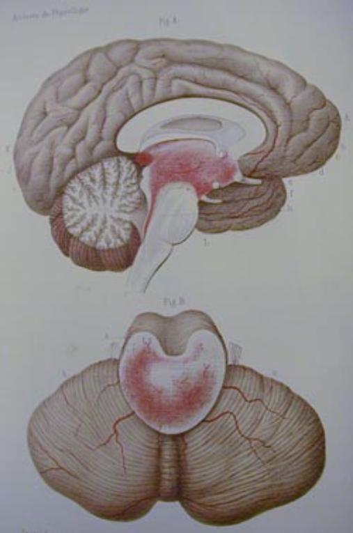
Hemmorrhagic
damage of the brainstem caused
by scalding of the airways,
drawing from Gayet (1875).
|

1 - Predilection sites
Decreased Purkinje cell density and abnormalities of
the cerebellar vermis and inferior olives have been
among the most consistent findings in the brains of
people with autism (Williams et al. 1980, Ritvo et al.
1986, Courchesne 1995, Hashimoto et al. 1995,
Bailey et al. 1998, Kemper & Bauman 1998). Kemper
and Bauman also described anomalies of cell size and
distribution in the limbic system, which included the
mammillary bodies. These are brain structures long
recognized as sites affected in Wernicke’s
encephalopathy. That autistic behaviors have been
observed in children exposed to alcohol during
gestation further suggests that impairment of function
in the brain areas affected in Wernicke’s
encephalopathy should be investigated as sites of
possible brain dysfunction in autism.
The bilaterally symmetric patterns of brainstem
damage seen in thiamine deficiency, Leigh syndrome,
and after resuscitation from asphyxia are all variants
of Wernicke’s encephalopathy. Wernicke (1881a)
described this kind of damage in a young woman who
ingested sulfuric acid and two alcoholic men.
Wernicke also cited a case reported by Gayet (1875)
who described similar clinical and pathological
features in a man injured in a boiler explosion. Figure
21 is a color plate from the article by Gayet, a graphic
illustration of the hemorrhagic nature of the damage
he observed in the brainstem. Brain damage was
more extensive in Gayet’s patient than in Wernicke’s
cases, with bilateral involvement of the superior
colliculi.
Kant (1933) found damage of the superior colliculi in
a few of the brains he examined, but emphasized the
more usual and severe involvement of the inferior
colliculi. Gayet’s patient survived five months after
the accident and scalding of the airway may have lead
to chronic hypoxia which in turn would lead to more
widespread damage of the brain. Wernicke’s
alcoholic patients survived less than two weeks after
episodes of acute intoxication; the victim of sulfuric
acid poisoning survived a little more than two months.
Discussions of Wernicke's encephalopathy often
involve perplexity over the confinement of damage to
the brainstem. The autoradiographic techniques for
measuring blood flow and glucose uptake in fact
provide an explanation for the predilection sites of
alcohol intoxication, thiamine deficiency, and other
causes of Wernicke-like patterns of brain damage.
The brainstem nuclei of high blood flow and high
metabolic rate are susceptible to impaired function if
not outright damage.
It should no longer be a mystery that the brainstem
rather than cortical centers of the higher faculties are
most distinctly involved. How higher cognitive
functions become impaired in alcoholism deserves
investigation. The association tracts of the cerebral
cortex do not operate independently of inputs from
sensory pathways in the brainstem. Cortical damage
may not be visible, but function is clearly impaired.
- Williams RS et al. (1980)
Autism and mental retardation:
Neuropathologic studies
performed in four retarded
persons with autistic behavior.
- Ritvo ER et al. (1986) Lower
Purkinje cell counts in the
cerebella of four autistic
subjects: initial findings of the
UCLA-NSAC Autopsy
Research Report.
- Courchesne E (1995) New
evidence of cerebellar and
brainstem hypoplasia in
autistic infants, children and
adolescents: the MR imaging
study by Hashimoto and
colleagues.
- Hashimoto T et al. (1995)
Development of the brainstem
and cerebellum in autistic
patients.
- Bailey A et al. (1998) A
clinicopathological study of
autism.
- Kemper TL, Bauman M (1998).
Neuropathology of infantile
autism..
- Wernicke C (1881) Die acute,
haemorrhagische
Poliencephalitis superior.
- Brody IA, Wilkins RH. (1968)
Wernicke's encephalopathy.
- Gayet M (1875) Affection
encéphalique (encéphalite
diffuse probable) localisée aux
étages supérieurs des
pédoncules cérébraux et aux
couches optiques, ainsi qu’au
plancher du quatrième
ventricule et aux parois
latérales du troisième.
- Kant F (1933) Die
Pseudoencephalitis Wernicke
der Alkoholiker. (polio-
encephalitis haemorrhagica
superior acuta).
- Williams RS, Hauser S, Purpura DP, deLong GR, Swisher CN (1980) Autism and mental
retardation: Neuropathologic studies performed in four retarded persons with autistic
behavior. Archives of Neurology 37:748-753.
- Ritvo ER, Freeman BJ, Scheibel AB, Duong T, Robinson H, Guthrie D, Ritvo A (1986) Lower
Purkinje cell counts in the cerebella of four autistic subjects: initial findings of the UCLA-NSAC
Autopsy Research Report. American Journal of Psychiatry 143:862-6
- Courchesne E (1995) New evidence of cerebellar and brainstem hypoplasia in autistic infants,
children and adolescents: the MR imaging study by Hashimoto and colleagues. Journal of
Autism and Developmental Disorders25:19-22.
- Hashimoto T, Tayama M, Murakawa K, Yoshimoto T, Miyazaki M, Harada M, Kuroda Y (1995)
Development of the brainstem and cerebellum in autistic patients. Journal of Autism and
Developmental Disorders 25:1-18.
- Bailey A, Luthert P, Dean A, Harding B, Janota I, Montgomery M, Rutter M, Lantos P (1998) A
clinicopathological study of autism. Brain 121:889-905.
- Kemper TL, Bauman M (1998). Neuropathology of infantile autism. Journal of Neuropathology
fcand Experimental Neurology 57:645-652.
- Wernicke C (1881a) Die acute, haemorrhagische Poliencephalitis superior. Lehrbuch der
Gehirnkrankheiten für Ärzte und Studirende,Band II. Kassel: Theodor Fischer, pp 229-242.
- Brody IA, Wilkins RH. (1968) Wernicke's encephalopathy. Archives of Neurology. 19:228-32.
- Gayet M (1875) Affection encéphalique (encéphalite diffuse probable) localisée aux étages
supérieurs des pédoncules cérébraux et aux couches optiques, ainsi qu’au plancher du
quatrième ventricule et aux parois latérales du troisième. Archives de physiologie normale et
pathologique série 2, 2:23-351.
- Kant F (1933) Die Pseudoencephalitis Wernicke der Alkoholiker. (polio-encephalitis
haemorrhagica superior acuta). Archiv für Psychiatrie und Nervenkrankheiten 98:702-768.
Figure xx: Hemorrhagic damage of the brainstem caused by scalding of the airways,
drawing from Gayet (1875).