2 - Wernicke's (alcoholic) encephalopathy
Reports of autistic behaviors among children exposed
during gestation to alcohol and other drugs began to
appear in the 1990s [1-3]. The finding of autism
among children with fetal alcohol syndrome should be
considered alongside the finding of autism in children
exposed to rubella infection during pregnancy [45.
Most important, the question should be raised as to
what areas of the brain are affected both by infections
and alcohol or other toxic substances, especially
during gestation, and how this might lead to autism in
some cases. Autism has also been found in children
of mothers taking valproic acid (Depakote) during
pregnancy [4, 6]. Autism has also been reported in
some victims of severe deformities caused by
maternal use of thalidomide during pregnancy [7].
It has been known for over a century how the brain is
affected by alcohol [8]. Wernicke (1881) first reported
the characteristic pattern of bilaterally symmetric
hemorragic lesions within the brainstem caused by
chronic alcohol intoxication. This pattern of selective
brainstem damage now bears the name Wernicke's
encephalopathy and its association with alcoholism
has been confirmed many times over. This pattern of
damage can be compared to the ischemic lesions
caused by brief total asphyxia at birth [9]. The inferior
colliculi are affected in most cases whether by
alcoholism or asphyxia, but the most predictable
lesions caused by alcohol abuse are in the mammillary
bodies [10, 11].
.
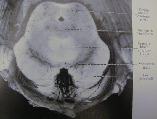
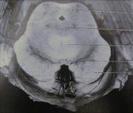
Figure 11 is from a paper by Kant (1933) and shows
petechial (pinpoint-size hemorrhagic spots) in the
inferior colliculi and surrounding areas of the midbrain,
which are characteristic of the damage in Wernicke's
encephalopathy [12]. The early papers, written in
German, describe the damage as small "flea-bite" size
hemorrhages that result from engorgement then
bursting of capillary vessels, similar to the "whiskey
nose" of many alcoholics.
The mammillary bodies are among the brainstem
nuclei of high metabolic rate, slightly less active than
the inferior colliculi (see tables 3 and 5, which are
discussed further in chapter xx). Protective
biofeedback mechanisms that spare the inferior
colliculi may make the mammillary bodies more
vulnerable to damage, just as motor areas become
more susceptible to damage in hypoxic situations.
The mammillary bodies are part of the limbic system,
in which Kemper and Bauman (1998) found signs of
disrupted prenatal development in brains from some
autistic individuals [13].
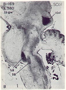
Figure 10a shows myelin stain in a midline (or sagittal)
section of the brainstem of a human fetus at 25
gestational weeks (gw) from Yakovlev and Lecours
(1967); this can be compared with the transverse
section in figure 10b [14]. Figure 10a shows the
greater degree of myelination in the superior olive
(SOl) and trapezoid body (TzB) of the auditory
pathway compared for example with the lesser degree
of myelination in the superior colliculus (SCol) of the
visual system.
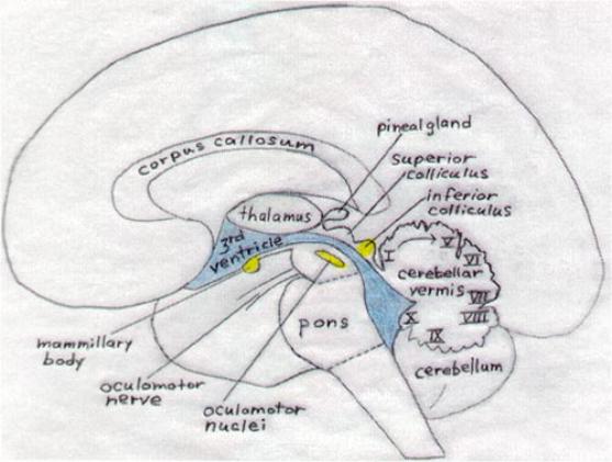
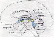
Figure 12 is a diagram of a sagittal view showing the
location of brainstem structures of high metabolic rate
that are vulnerable to damage from alcohol
intoxication and other factors that impair aerobic
metabolism. Structures shown in figure 9 can be
compared to degrees of prenatal myelination of those
in figure 6a. Greater detail and orientation can be
found by studying diagrams (transverse, sagittal, and
coronal) given in textbooks of neuroanatomy, like that
of Nolte and Angevine [15].
- Nanson JL (1992) Autism in
fetal alcohol syndrome: a
report of six cases.
- Harris SR et al. (1995) Autistic
behaviors in offspring of
mothers abusing alcohol and
other drugs: a series of case
reports.
- Aronson M et al. (1997)
Attention deficits and autistic
spectrum problems in children
exposed to alcohol during
gestation: a follow-up study.
- Chess S (1971) Autism in
children with congenital
rubella.
- Christianson AL et al.(1994)
Fetal valproate syndrome:
clinical and neuro-
developmental features in two
sibling pairs.
- Williams G et al.(2001) Fetal
valproate syndrome and
autism: additional evidence of
an association.
- Stromland K et al. (1994)
Autism in thalidomide
embryopathy: a population
study.
- Wernicke C (1881a) Die acute,
haemorrhagische
Poliencephalitis superior.
- Windle WF (1969a) Brain
damage by asphyxia at birth.
- Torvik A (1987) Topographic
distribution and severity of
brain lesions in Wernicke's
encephalopathy.
- Victor M, Adams RD, Collins
GH (1989) The Wernicke-
Korsakoff syndrome and
related neurologic disorders
due to alcoholism and
malnutrition, 2nd ed.
- Kant F (1933) Die
Pseudoencephalitis Wernicke
der Alkoholiker. (polio-
encephalitis haemorrhagica
superior acuta)..
- Kemper TL, Bauman M (1998).
Neuropathology of infantile
autism.
- Yakovlev PI and Lecours A-R
(1967) The myelogenetic
cycles of regional maturation
of the brain.
- Nolte J, Angevine JB (1995)
The Human Brain, in
Photographs and Diagrams.
Pinpoint hemorrhages
in the inferior colliculi from
alcohol intoxication
Figure 12 - Diagram showing brainstem sites affected in Wernicke's encephalopathy
|
Figure 10 - From Yakovlev & Lecours (1967)
showing prominent myelination in the inferior
colliculus (ICOL) at 25 gestational weeks
(below) and 29 gestational weeks (right).
|
From Yakovlev & Lecours (1967) with permission from Blackwell Scientific Publishers
|
LLm - lateral lemniscus (auditory)
Mlm - medial lemniscus (motor)
|
ICol - inferior colliculus (auditory)
Scol - superior colliculus (visual)
TzB - trapezoid body (auditory)
SOl -superior olive (auditory)
|
Figure 11 - From Kant (1933). Corpus quadrigeminum posterior, another name for
|
|
\/
- Nanson JL. Autism in fetal alcohol syndrome: a report of six cases. Alcohol Clin Exp
Res. 1992 Jun;16(3):558-65.
- Harris SR, MacKay LL, Osborn JA (1995) Autistic behaviors in offspring of mothers
abusing alcohol and other drugs: a series of case reports. Alcoholism, Clinical and
Experimental Research 19:660-5
- Aronson M, Hagberg B, Gillberg C (1997) Attention deficits and autistic spectrum
problems in children exposed to alcohol during gestation: a follow-up study.
Developmental Medicine and Child Neurology 39:583-7.
- Chess S (1971) Autism in children with congenital rubella. J Autism Child Schizophr.
1971 Jan-Mar;1(1):33-47.
- Christianson AL, Chesler N, and Kromberg JGR (1994) Fetal valproate syndrome:
clinical and neuro-developmental features in two sibling pairs. Developmental Medicine
and Child Neurology 36:357-369.
- Williams G, King J, Cunningham M, Stephan M, Kerr B, Hersh JH. (2001) Fetal valproate
syndrome and autism: additional evidence of an association. Developmental Medicine
and Child Neurology 43:202-206.
- Stromland K, Nordin V, Miller M, Akerstrom B, and Gillberg C (1994) Autism in
thalidomide embryopathy: a population study. Developmental Medicine and Child
Neurology 36:351-356.
- Wernicke C (1881a) Die acute, haemorrhagische Poliencephalitis superior. Lehrbuch
der Gehirnkrankheiten für Ärzte und Studirende,Band II. Kassel: Theodor Fischer, pp
229-242.
- Windle WF (1969a) Brain damage by asphyxia at birth. Scientific American 221(#4):76-
84.
- Torvik A (1987) Topographic distribution and severity of brain lesions in Wernicke's
encephalopathy. Clinical Neuropathology 6:25-29.
- Victor M, Adams RD, Collins GH (1989) The Wernicke-Korsakoff syndrome and related
neurologic disorders due to alcoholism and malnutrition, 2nd ed, Contemporary
Neurology Series v30. Philadelphia, PA : F.A. Davis Co.
- Kant F (1933) Die Pseudoencephalitis Wernicke der Alkoholiker. (polio-encephalitis
haemorrhagica superior acuta). Archiv für Psychiatrie und Nervenkrankheiten 98:702-
768.
- Kemper TL, Bauman M (1998). Neuropathology of infantile autism. Journal of
Neuropathology fcand Experimental Neurology 57:645-652 .
- Yakovlev PI, Lecours A-R (1967) The myelogenetic cycles of regional maturation of the
brain. In A. Minkowski (Ed.), Regional Development of the Brain in Early Life (pp. 3-70).
Oxford: Blackwell Scientific Publications.
- Nolte J and Angevine JB (1995) The Human Brain, in Photographs and Diagrams.
Mosby, St. Louis.
: Brainstem sites
affected in Wernicke's
Encephalopathy
Early maturation and function in
the brainstem auditory pathway
Highest blood flow in the brain
is to the inferior collicui
Highest glucose uptake in the
brain is in the inferior colliculi
Pinpoint hemorrhages from chronic alcohol intoxication are evident in the inferior colliculi.
Table 3: Cerebral blood flow data in cats, using a
radiographic tracer (from Landau et al. 1955)
|
Brain Structure
Brain System
Flow Rate
(cc/gm/min)
Auditory
Auditory
Visual
Visual
Subcortical motor
1.80
1.38
1.30
1.25
1.22
1.21
1.15
1.10
1.03
0.88
0.87
0.24
0.23
0.14
Inferior colliculus
Sensory-motor cortex
Auditory cortex
Visual cortex
Medial geniculate
Lateral geniculste
Superior colliculus
Caudate nucleus
Thalamus
Association cortex
Cerebellar nuclei
Cerebellar white matter
Cerebral white matter
Spinal cord white matter
TABLE 5: Glucose utilization (from Sokoloff 1981)
|
Uptake of radioactive deoxyglucose in monkeys and rats
|
Brain structure
Brain system
Albino rat
(sd 2-7)
Monkey
(sd 1-4)
auditory
auditory
auditory
limbic
visual
subcortical motor
auditory
visual
limbic
197
162
128
131
133
107
121
95
116
110
113
100
120
96
79
57
37
103
79
66
65
63
59
57
55
54
52
51
45
44
39
39
31
12
Inferior colliculus
Auditory cortex
Vestibular nucleus
Medial geniculate
Superior olive
Visual cortex
Mammillary body
Superior colliculus
Thalamus, lateral
Caudate-putamen
Cochlear nucleus
Cerebellar nuclei
Sensorimotor cortex
Lateral geniculate
Hippocampus
Cerebellar cortex
Cerebellar white